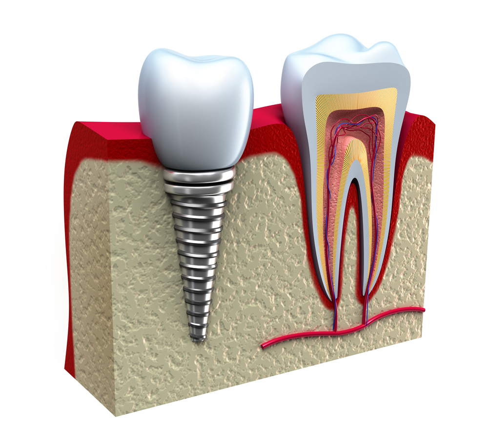
Core: indications, techniques and clinical steps

INDICATIONS
In teeth with some degree of destruction, requiring prosthetic treatment
Goals
Recover the anatomical characteristics of the clinical crown, giving the prepared tooth biomechanical conditions to maintain the prosthesis in function for a reasonable period.
TECHNIQUES
They vary according to the degree of coronary destruction and whether the tooth has pulp vitality.
1- Pulped Teeth (Fillers)
The amount of coronal structure remaining after tooth preparation should be analyzed. After this preparation and depending on the amount of coronal structure remaining, it is easier to decide whether endodontic treatment is required.
Basic rule: If there is approximately half of the coronary structure and preferably surrounding the cervical third of the tooth (region responsible for frictional retention of the crown) the rest of the crown can be restored with filler material (composite resins, glass ionomers and combination of both, which is called compomers), using intradental threaded pins to give greater retention.
The choice of filler materials is due to the properties (elasticity) being like the properties of dentin and the fact that it has an excellent adhesion capacity.
Note: If there is not enough dental structure to resist chewing forces to avoid the risk of fractures, we should perform endodontic treatment. We should, as much as possible, avoid devitalization of a tooth, as placement of the intra canal metal pin tends to weaken the remaining root's dental structure, making it more susceptible to fractures. There is also a risk of trepanning during the work performed.
2- Pulped Teeth (Intra-root fused cores)
Restoration with fused nuclei due to major coronary destruction, where the coronary remnant is not enough to provide structural resistance to the filler material.
- Preparation of coronary remnant:
The preparation should be performed following the characteristics of the type of prosthesis indicated. The temporary cement contained in the pulp chamber is removed until the mouth of the duct. The pulp chamber retentions are removed.
The prepared crown walls should have a support base for the core with a thickness of 01 mm. It is on this basis that forces are directed to the root of the tooth, minimizing stress.
When there is not enough coronary structure to provide this support base, a box inside the root approximately 2mm deep should be prepared to create a core support base that will direct forces vertically, reducing tensions in the walls. root sides. These boxes can only be made if the root has enough structure.
- Conduit preparation:
There are 4 factors that we must analyze to provide adequate retention of the intraradicular nucleus: Length, Wall Slope, Diameter and Surface Characteristic.
First factor: Length.
Must be equal to or larger than the clinical crown. As a rule, the length of the intraradicular pin should reach 2/3 of the total length of the dental remnant.
“The correct length of the core inside the root is synonymous with longevity of the prosthesis.” The proper length of the pin inside the root provides a more even distribution of occlusal forces over the entire root surface, reducing the possibility of concentration. stress in certain areas and consequently fractures.
The length of the pin should be analyzed and determined by a periapical radiograph after the preparation of the coronary portion, taking into account the minimum amount of 4mm of obturator material that should be left in the apical region of the root canal to ensure an effective seal in this region. .
Note: Short core favors stress concentration in certain areas, causing root fracture.
Partial Endodontic Treatment:
When the obturator material has not reached the desired level, we should consider the treatment time and the presence of periapical lesion.
- Partial endodontic treatment with lesion: conduit retreatment is indicated.
- Partial endodontic treatment with no lesion: we should consider the treatment time. If it was made at least 5 years ago we can put the core, keeping the remaining material obturator.
Note: If the prepared proportion of the conduit is not considered adequate to establish the length of the nucleus, the retreatment of the canal is indicated, regardless of time and absence of lesion.
- Teeth filled with silver cones: retreatment is recommended to receive the fused cores while maintaining apical sealing.
Second factor: Wall Slope.
- Sloping walls: less retention than parallel walls. Forces the surrounding walls and can cause fractures.
- Parallel walls: ideal format !!! It should be similar to the inclination of the conduit itself that has been widened by endodontic treatment and will have a greater increase in wear in the most apical region for intraradicular nucleus placement until it has adequate length and diameter.
Note: If the duct has steeply sloping walls, the practitioner should increase the length of the intraradicular pin to achieve some form of parallelism to the walls near the apical region and make the most of the remaining coronal portion that will aid in retention and minimize the distribution of efforts at the root of the tooth.
Tooth with very wide duct and very thin walls - we can use “stowed” cores to protect the root. This type of nucleus seeks intraradicular retention and protects the thin walls of the root remnant.
Third Factor: Diameter.
The pin diameter should be up to 1/3 of the total root diameter. The dentin thickness should be greater on the buccal surface of the upper anterior teeth because the incidence of force is greater in this direction. Clinically, the diameter of the pin should be determined by radiography comparing the diameter of the drill with the diameter of the duct. For the metal used to have satisfactory resistance, it must be at least 1 mm in diameter at its apical end.
Fourth factor: Surface Feature
To increase retention of cast cores that have smooth surfaces, such surfaces should be turned into rough or uneven regions prior to cementation with the aid of drills or blasting with aluminum oxide.
CLINICAL STEPS
1- Removal of obturator material and conduit preparation
The obturator material should first be removed with heated Rhein tips to the preset length.
If it is not possible to use this instrument, the desired amount of Peeso, Gates or Largo drill bits of appropriate diameter to the duct must be removed, coupled with a penetration guide.
During the use of the drill we should always follow the extension of the conduit visualizing the obturator material and being careful not to risk trepanning the root.
The obturator material should be removed to a certain extent as a minimum of 04 millimeters of obturator material must be left at the root of the root to ensure effective sealing in this region.
Note 01: For multi-root teeth with parallel ducts, the duct preparation need not be the same length. Only the largest diameter conduit is carried to its maximum length of 2/3 and the other only to half of the total length of the remaining crown-root. As the conduits are parallel we can have two pins joined by the same base acting as an anti-rotational device, thus, it is not necessary to widen and oval the conduits, seeking to reach the minimum diameter of 01 mm for the alloy maintain its resistance characteristics.
Note 02: Superior premolars present divergence of the roots and, therefore, should have their bulkier conduit prepared to 2/3 extension and the other conduit partially prepared only for the purpose of stability, acting as an anti-rotational device.
Note 03: upper multiradicular teeth with divergent remaining coronary conduits, the palatine conduit is prepared up to 2/3 of its extension and one of the buccal conduits up to its half (the largest of them) and the other only part of its prepared mouth.
Note 04: Multiradicular teeth with divergent ducts in the absence of the coronary remnant completely, the three divergent ducts should be prepared and the resulting nucleus should be made in three distinct parts.
Note 05: Lower molars have mesial root with parallel or slightly divergent conduits and rarely require division of the nucleus into more than two segments as they may become parallel through the preparation.
2- Confection of the core
There are two techniques for core making:
- Direct: Conduit is molded and the coronary part is carved directly into the mouth.
- Indirect: requires shaping of the conduits and remaining coronary portion with elastomers, obtaining a model on which the cores are sculpted in the laboratory. This technique is indicated when it is necessary to make cores for several teeth or for teeth with divergent roots.
Direct Technique - Unirradicular Tooth
1- A rod of acrylic resin is prepared that adapts to the diameter and length of the prepared conduit and extends 1 centimeter beyond the remaining crown. It is necessary for the rod to reach the apical portion of the prepared duct and that there is space between it and the axial walls to facilitate molding of the duralay resin duct.
2- Lubricate the conduit and the coronary portion with the aid of a Peeso (or similar) drill wrapped in cotton.
3- The conduit is molded by lifting the resin prepared with a Centrix probe, brush or syringe inside and wrapping the stick inserted in it, until checking if its full extent has been reached. The remaining material is used to make the coronary portion of the nucleus.
Note 01: For teeth with two parallel ducts, the ducts are individually molded and, after polymerization of the resin, are joined in the region of the pulp chamber.
Note 02: During polymerization of the acrylic resin, the stick should be removed and inserted several times into the duct to prevent the core from being trapped by the retention left during duct preparation. After polymerization of the resin, the molded pin is checked for fidelity and the rod is cut at the occlusal or incisal level to continue the preparation of the coronary portion using the drills and sanding discs.
Note 03: The coronary part of the nucleus should only complement the missing tooth structure, giving it the shape and characteristics of a prepared tooth.
4- The metal alloy used in the foundry must be strong enough not to deform under chewing forces. The noble copper-aluminum alloy is the most used because it is low cost, but there are also noble alloys such as: type III and type IV, based on palladium silver.
5- After adaptation the root portion of the core should be blasted with aluminum oxide.
6- Before cementing it is necessary to clean the flue with absolute alcohol.
7 - Cementation may be performed with zinc phosphate or glass ionomer. Take the brush with a small amount of cement around the core.
Direct Technique - Multiradicular Tooth
Nucleus in teeth with divergent roots can also be made by direct technique, either by molding the ducts with resin or by using prefabricated systems.
1- Molding the ducts with resin: the procedures for preparing the ducts and making the cores follow the same principles described above.
Note: Another way to obtain cores by direct technique in teeth with divergent ducts is to initially make the pin of the larger volume canal that will pass through the coronary portion of the nucleus.
2- Shaping the ducts with prefabricated pins (Parapost System): This system has prefabricated metal and plastic pins, with parallel and serrated walls in various diameters and their respective drills.
Indirect technique
We fit in each duct an orthodontic wire or paperclip, slightly longer than the duct and slightly slack all around the duct walls. The wires should have at their end, facing the occlusal, a restraint system that can be made with low fusion shield. To bring the impression material to the ducts use a drill manually or coupled to a counter angle, rotating the engine at low speed. The metallic wires are also wrapped with the material, placed in their respective conduits and then, with an appropriate syringe, the coronary part is molded, completely enveloping the metallic wires that are in position.
Note 01: Any elastomer may be employed for duct shaping provided it provides the laboratory technician with an accurate and reliable model for obtaining split or multiple cores.
Note 02: To make the working model, cast the mold with plaster type IV.
Note 03: Models must be mounted on DENTFLEX articulators to maintain proper antagonist tooth relationships.
Core confection
Once fused, the first core part is adapted to the working model and the face finish that will contact the other core part is finished. Then the second part is made which, after being cast and adapted to the model, is adjusted to the tooth.
Cementing is performed with the introduction of the first core part (female part of the socket), followed by the second part (male).
Restore with prefabricated cores
When the element to be restored has endodontic treatment and maintains a considerable part of the clinical crown after tooth preparation, the placement of a prefabricated pin in the root canal is indicated to increase the resistance of the filling material. These pins can be smooth, serrated or threaded and differ in their surface morphology inside the root canal.
Note: Threaded studs have greater retention capacity, but unfortunately generate more stress on root canal walls than cemented ones. When indicated for use, unscrew 1/4 turn after final insertion into the duct to minimize dentin stresses. Preferably, the prefabricated pins should be cemented and juxtaposed in the root canal passively.
There are several makes and types of these prefabricated pins, and the choice should be determined based on the relationship: duct diameter / pin length. The duct diameter must be compatible with the pin diameter; which should be selected from the comparison between its diameter and the conduit space by radiography. The presence of a thickness of 2 to 3 millimeters in the remaining portion of the root significantly increases its fracture resistance.
Note 01: The conduit is prepared at 2/3 the size of the tooth, from its coronary portion to the apex. When the tooth has bone loss, the pin length should be equivalent to half of the involved root bone support.
Note 02: When it is a posterior tooth with two or more roots, consider whether this tooth will receive an isolated crown or whether it will be used as the abutment tooth of a fixed prosthesis and how long it is.
For isolated elements and even for 3-element fixed prosthesis, considering that the tooth still has coronary remnant, there is no need for all roots to receive metal pins. We should choose only the largest diameter root.
Removal of the obturator material should be performed initially with the Rhein tips heated to the pre-established length and then the conduit should be widened with the drills that accompany the metal pins, or with the Peeso, Gates or Largo drills. with diameter compatible with the conduit.
Note: The pin is cemented with zinc phosphate or glass ionomer and the filler core is made of composite resin or compomer.
Core confection with reuse of existing prosthesis
The main cause of failure in fixed prosthesis is dental caries and, therefore, cemented fixed prostheses for some time may have decayed abutment teeth. In these cases, and as long as the prosthesis can remain in the mouth, the nucleus can be made in a conventional manner, and its coronary portion is obtained by molding the interior of the crown.
1- Tooth that required endodontic treatment due to carious process.
2- Conduits are prepared and molded leaving a small projection of the resin to the occlusal.
3- The entire inner surface of the crown is slightly worn, including the cervical region, to eliminate possible retentive areas.
4- The resin is prepared and taken to the region corresponding to the cervical terminus and inside the crown that is positioned over the tooth, taking care to evaluate the occlusion.
5- After resin polymerization, the coronary portion of the nucleus is evaluated, and the cervical region is finished.
6- After the resin core is cast, it is cemented in the duct and then the crown is cemented.
Source: Profissão Dentista. Available at: https://profissaodentista.com/2017/03/06/nucleos-indicacoes-tecnicas-etapas-clinicas/. Access on: 09/01/2020.
